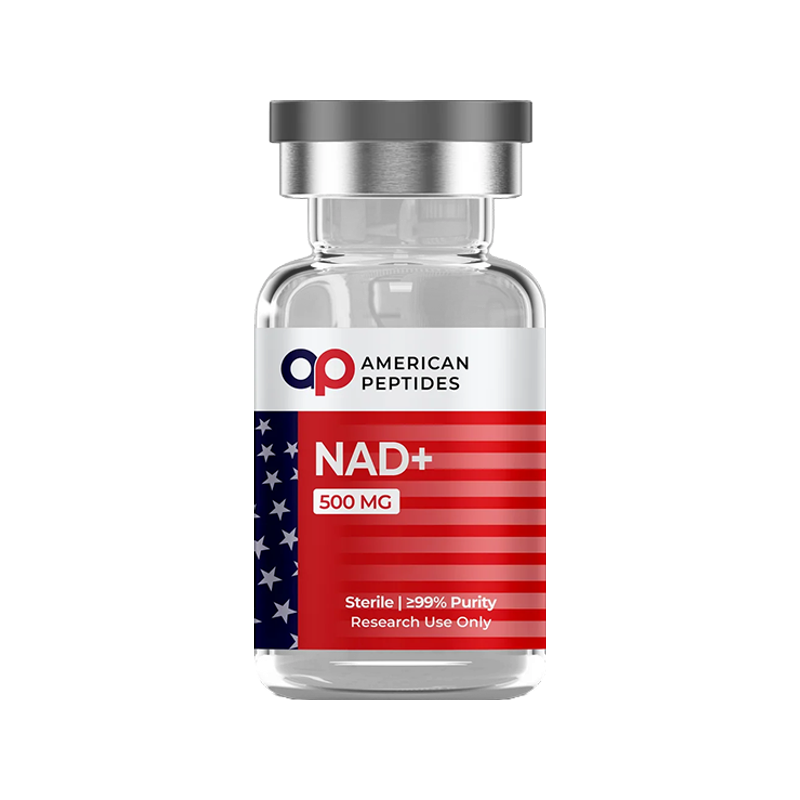

Nicotinamide adenine dinucleotide — NAD+ in its oxidized form, NADH in its reduced form — is not a peptide. It is a dinucleotide coenzyme present in every living cell, and it sits in a different chemical category from everything else in a research-peptide catalogue. This summary builds on our short NAD+ glossary entry and goes deeper: how the molecule is made and consumed, what cell and tissue-sample studies report about its behaviour, what rodent models have shown about why its levels fall with age, and where the human evidence actually stops.
Research-use-only context. This article summarizes published third-party scientific literature — the large majority of it conducted in cultured cells or animal models. It is not medical advice, not a therapeutic or performance claim, and not a usage guide. American Peptides products are sold strictly for in vitro laboratory research and are not for human or veterinary use.
What NAD+ is
Structurally, NAD+ is two nucleotides joined through their phosphate groups: one carries adenine, the other carries nicotinamide. There are no amino acids and no peptide bonds anywhere in the molecule, so it is not a peptide by any definition. Its defining chemistry is the nicotinamide ring, which accepts a hydride (two electrons and a proton) to become NADH and gives it back again; that reversible exchange is what makes NAD+ a redox coenzyme. A 2021 review in Nature Reviews Molecular Cell Biology describes it both as a coenzyme for redox reactions and as an essential cofactor for non-redox enzymes that consume it outright — sirtuins, CD38 and poly(ADP-ribose) polymerases (PARPs).1 Those two roles, electron carrier and enzyme substrate, frame everything that follows.
Cells make NAD+ by three routes, and the research literature on NAD+ decline is largely a literature about these routes. The de novo pathway builds the pyridine ring from the amino acid tryptophan over several enzymatic steps. The Preiss–Handler pathway starts from nicotinic acid (niacin), converting it to nicotinic acid mononucleotide and onward to NAD+. The salvage pathway recycles nicotinamide — the fragment released every time a sirtuin, PARP or CD38 cleaves NAD+ — back into NAD+. Its first step is catalysed by nicotinamide phosphoribosyltransferase (NAMPT), which converts nicotinamide to nicotinamide mononucleotide (NMN); a 2011 Cell Metabolism paper describes NAMPT as the rate-limiting enzyme in mammalian NAD+ biosynthesis and NMN as the product of its reaction.8
The two precursors most often discussed alongside NAD+ are best understood as chemistry. Nicotinamide riboside (NR) is a nucleoside: the nicotinamide ring attached to a ribose sugar. NMN is the same nucleoside with a phosphate added, one adenylation step away from NAD+ itself. The 2004 Cell paper that established NR as an NAD+ precursor reported that the accepted view — that all eukaryotic NAD+ synthesis flows through nicotinic acid mononucleotide — did not hold in yeast, identified nicotinamide riboside kinases (NRKs) from yeast and humans that phosphorylate NR specifically, described this as a Preiss–Handler-independent route, and reported NR as a natural constituent of milk.3 NR therefore enters the pathway at the NMN step through NRK, and nicotinamide enters it through NAMPT; both converge on NAD+.
What the research reports
Redox chemistry and the NAD+/NADH ratio
Because NAD+ and NADH interconvert constantly in glycolysis, the citric-acid cycle and mitochondrial respiration, researchers care about the ratio between the two forms as much as the absolute amount. Standard assays require extracting and destroying the sample, so a 2015 PNAS study that quantified NAD+, NADH and the NAD+/NADH redox potential non-invasively by magnetic resonance in the intact human brain was a methodological step. In healthy volunteers it reported an age-dependent increase in intracellular NADH alongside age-dependent reductions in NAD+, total NAD and the NAD+/NADH redox potential, which the authors interpret as altered NAD homeostasis accompanying normal aging.5 It is an observational measurement in a modest cohort, but a direct in-situ readout rather than a rodent extrapolation.
NAD+-consuming enzymes: sirtuins, PARPs and CD38
Redox cycling does not use NAD+ up; the same molecule is oxidized and reduced thousands of times. The enzymes that actually consume it cleave the molecule at the glycosidic bond and release nicotinamide. The Nature Reviews Molecular Cell Biology review groups them into three families: sirtuins (NAD+-dependent deacylases linked to chromatin remodelling and metabolic regulation), PARPs (DNA-repair enzymes that build poly(ADP-ribose) chains from NAD+), and CD38 (an ecto-enzyme that hydrolyses NAD+). Through them, the review notes, NAD+ availability can influence DNA repair, chromatin state, cellular senescence and immune-cell function.1 The 2012 PLoS One human-tissue study makes the competitive point plainly: in genomic DNA, NAD+ is the sole substrate for PARP, and PARP shares that pool with the sirtuins, so rising PARP activity in response to DNA damage leaves less NAD+ for sirtuins.4 The 2016 Cell Metabolism CD38 paper adds a wrinkle: CD38 was identified as the main enzyme degrading the precursor NMN in vivo, so it acts upstream of NAD+ synthesis as well as downstream of it.6
Tissue NAD+ declines with age in rodents and in human samples
The most consistent finding in this field is that tissue NAD+ falls with age. The Nature Reviews authors describe a gradual decline in multiple model organisms, including rodents and humans, and are candid that the mechanisms regulating those levels are not fully understood.1 The 2011 Cell Metabolism study reported that NAD+ and NAMPT levels decreased significantly in multiple organs of aging mice.8 In humans, the clearest tissue-sample evidence is the 2012 PLoS One study of pelvic skin from 49 donors ranging from newborns to age 77. NAD+ content showed a strong negative correlation with age in both sexes; in males, PARP activity rose with age and correlated inversely with tissue NAD+, and SIRT1 activity declined, while those associations were weaker or absent in females.4 Together with the brain magnetic-resonance work,5 these are the human observations behind the claim that NAD+ declines with age. They are consistent, but their scale is one skin-sample series and one imaging cohort.
Animal-model findings: CD38 and NAMPT
Why NAD+ falls with age has been studied mostly in mice, and two threads dominate. The first is NAMPT and supply. The 2011 Cell Metabolism paper reported that NAMPT-mediated NAD+ biosynthesis was severely compromised in metabolic organs of mice on a high-fat diet, and that supplying the NAMPT product NMN restored NAD+ levels and improved glucose tolerance in those animals, partly through SIRT1 activation.8 The second thread is CD38 and demand. The 2016 Cell Metabolism study reported that CD38 expression and activity increase with aging in mice and that CD38 is required for the age-related NAD decline and associated mitochondrial dysfunction, acting at least in part through SIRT3.6 A 2020 Nature Metabolism paper asked where that extra CD38 comes from and reported that pro-inflammatory M1-like macrophages — but not naive or M2 macrophages — accumulate in visceral white adipose tissue and liver during aging, express high levels of CD38, and show enhanced CD38-dependent NADase activity; senescent cells accumulate in the same tissues, and the cytokines they secrete induce macrophages to proliferate and express CD38.7 Read together, these papers propose a mechanism: inflammation and senescence drive up an NAD+-consuming enzyme in tissue-resident immune cells while the salvage enzyme loses capacity. It is a coherent story, and it is a mouse story. Whether the same balance operates in aging human tissue has not been established.
The human-evidence gap
Three things need saying plainly. First, almost all human interventional literature concerns oral precursor supplementation trials, not NAD+ itself. The results have been mixed and modest. A 2024 placebo-controlled pilot in GeroScience, for example, randomised 20 older adults with mild cognitive impairment to NR or placebo and reported that blood NAD+ rose roughly 2.6-fold in the NR group while cognitive and psychometric measures remained stable throughout, with no between-group difference on the primary outcome. A change in cerebral blood flow was observed but, the authors note, would not have survived correction for multiple comparisons, and they call for a larger, longer trial.2 That pattern — a rise in a blood biomarker without a change in the functional endpoint — recurs across this literature.
Second, NAD+ the compound is a different chemical entity from the precursors used in those trials, with its own handling and stability profile in solution, and the trial findings do not transfer to it. Research-grade NAD+ supplied as a laboratory reagent has no human data of its own: no controlled trials, no outcome studies, nothing. It is characterised as a biochemical — purity, identity, redox state — and nothing more. Third, NAD+ has no FDA drug approval for any indication. Reviewers are explicit that much remains to be learnt about how NAD+ influences human aging biology, including whether restoring its levels in aging humans is safe and whether it does anything useful.1 That is the state of the evidence, and it is considerably more cautious than the marketing suggests.
The takeaway
NAD+ is a well-understood coenzyme with a rich mechanistic literature: a redox carrier that is also the shared substrate of sirtuins, PARPs and CD38, synthesised by three converging routes with NAMPT as the rate-limiting salvage step. Human tissue-sample and imaging studies report that it declines with age; mouse studies implicate rising CD38 in immune cells and falling NAMPT capacity. What the literature does not yet contain is human evidence that intervening on any of this changes a meaningful outcome, and the dinucleotide itself has essentially no human data at all. American Peptides supplies NAD+ strictly for in vitro research, as a reference compound for laboratories studying redox chemistry and NAD+-dependent enzymes. For the mitochondrial context in which most of this work is framed, see What is mitochondrial health?
Frequently Asked Questions
Is NAD+ a peptide?
No. NAD+ is a dinucleotide — two nucleotides (one adenine, one nicotinamide) linked through phosphate groups. It contains no amino acids and no peptide bonds.
How do cells make NAD+?
By three routes: de novo synthesis from tryptophan, the Preiss–Handler pathway from nicotinic acid, and the salvage pathway, in which NAMPT converts nicotinamide to NMN and NMN is converted to NAD+. Nicotinamide riboside enters at the NMN step through NRK enzymes, a route first described in 2004.
Why does NAD+ decline with age?
Human skin-sample and brain-imaging studies report lower NAD+ with increasing age. Mouse studies attribute the decline to rising activity of the NAD+-consuming enzyme CD38 in tissue-resident immune cells, driven by inflammation and senescent cells, together with reduced capacity of the NAMPT salvage pathway. Whether the same mechanism operates in humans has not been shown.
Is NAD+ approved by the FDA?
No. NAD+ has no FDA drug approval for any indication, and the human trial literature concerns oral precursor supplements rather than NAD+ itself. American Peptides supplies it strictly for in vitro research.
Citations
- Covarrubias AJ, et al. “NAD+ metabolism and its roles in cellular processes during ageing.” Nat Rev Mol Cell Biol. 2021;22(2):119–141. PubMed: PMID 33353981
- Orr ME, et al. “A randomized placebo-controlled trial of nicotinamide riboside in older adults with mild cognitive impairment.” Geroscience. 2024;46(1):665–682. PubMed: PMID 37994989
- Bieganowski P, Brenner C. “Discoveries of nicotinamide riboside as a nutrient and conserved NRK genes establish a Preiss-Handler independent route to NAD+ in fungi and humans.” Cell. 2004;117(4):495–502. PubMed: PMID 15137942
- Massudi H, et al. “Age-associated changes in oxidative stress and NAD+ metabolism in human tissue.” PLoS One. 2012;7(7):e42357. PubMed: PMID 22848760
- Zhu XH, et al. “In vivo NAD assay reveals the intracellular NAD contents and redox state in healthy human brain and their age dependences.” Proc Natl Acad Sci U S A. 2015;112(9):2876–2881. PubMed: PMID 25730862
- Camacho-Pereira J, et al. “CD38 Dictates Age-Related NAD Decline and Mitochondrial Dysfunction through an SIRT3-Dependent Mechanism.” Cell Metab. 2016;23(6):1127–1139. PubMed: PMID 27304511
- Covarrubias AJ, et al. “Senescent cells promote tissue NAD+ decline during ageing via the activation of CD38+ macrophages.” Nat Metab. 2020;2(11):1265–1283. PubMed: PMID 33199924
- Yoshino J, et al. “Nicotinamide mononucleotide, a key NAD+ intermediate, treats the pathophysiology of diet- and age-induced diabetes in mice.” Cell Metab. 2011;14(4):528–536. PubMed: PMID 21982712
Related reading
This article is for laboratory research reference only. American Peptides products are sold strictly for in vitro research. Not for human consumption.
Related research
Compliance Notice: American Peptides products are sold strictly for laboratory and academic research purposes only. They are not intended for human or veterinary consumption, diagnosis, treatment, or prevention of any disease. All content on this page is educational in nature and does not constitute medical advice or product claims. Researchers are responsible for handling these compounds in accordance with their institution’s safety protocols and applicable laws.