
Amino Acid Signaling and Receptor Biology: A Research Primer
What is receptor biology? Receptor biology is the study of how cells detect and respond to chemical signals from their environment. Cells use specialized proteins called receptors to recognize specific signaling molecules. When a signal binds its receptor, the receptor transduces the binding event into an intracellular response — a process called signal transduction. The four major receptor classes are G-protein-coupled receptors, receptor tyrosine kinases, ion-channel-linked receptors, and intracellular receptors.
What is signal transduction?
Cells live in a chemical world. They are surrounded by hormones, neurotransmitters, growth factors, cytokines, and locally released signaling peptides. To behave appropriately, a cell must recognize which signals are present and respond accordingly. The process of converting an extracellular chemical signal into an intracellular change is called signal transduction.
The architecture is conceptually simple. A signaling molecule (the ligand) is released by one cell. It diffuses through the extracellular space. It encounters a target cell whose surface bears a receptor specific for that ligand. The ligand binds the receptor. The receptor changes conformation. The changed conformation triggers downstream events inside the cell — sometimes a single phosphorylation, sometimes a cascade involving dozens of proteins, sometimes a change in gene expression that reshapes the cell over hours.
Every cellular response to its environment, from muscle contraction to insulin secretion to immune activation, ultimately depends on signal transduction. Receptors are the molecular machines that make it work.
The four major receptor classes
Receptors fall into four major structural classes, each transducing signals by a different mechanism:
   ┌────────────────────────────────────────────────────┐
   │  1. G-PROTEIN-COUPLED RECEPTORS (GPCRs)            │
   │     Seven-transmembrane proteins.                  │
   │     Couple to intracellular G proteins.            │
   │     ~800 in the human genome.                      │
   │                                                    │
   │  2. RECEPTOR TYROSINE KINASES (RTKs)               │
   │     Single-pass transmembrane proteins.            │
   │     Catalytic kinase activity in cytoplasmic tail. │
   │     ~58 in the human genome.                       │
   │                                                    │
   │  3. ION-CHANNEL-LINKED RECEPTORS                   │
   │     Multi-subunit transmembrane proteins.          │
   │     Ligand binding opens an ion channel.           │
   │     Examples: nicotinic ACh, GABA-A, NMDA.         │
   │                                                    │
   │  4. INTRACELLULAR (NUCLEAR) RECEPTORS              │
   │     Located in cytoplasm or nucleus.               │
   │     Bind lipophilic ligands that cross membrane.   │
   │     Function as ligand-activated transcription     │
   │     factors. Examples: steroid, thyroid, vitamin D.│
   └────────────────────────────────────────────────────┘
For peptide research, the first two classes are most relevant — most signaling peptides act through GPCRs or RTKs.
G-protein-coupled receptors (GPCRs) in depth
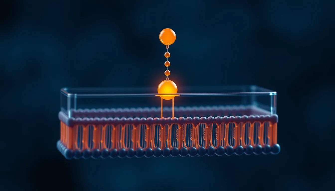
GPCRs are the largest family of cell-surface receptors in mammals — roughly 800 distinct GPCRs in the human genome, more than any other receptor class. They detect signals from hormones, neurotransmitters, light photons, odorant molecules, and tastant molecules. Every GPCR shares a common architecture: a single polypeptide chain that crosses the cell membrane seven times, with the N-terminus on the extracellular side and the C-terminus on the cytoplasmic side.
The signal transduction mechanism:
- Ligand binding to the extracellular face changes the receptor's conformation.
- The cytoplasmic face of the receptor activates an associated heterotrimeric G protein — a complex of α, β, and γ subunits.
- G protein activation involves the α subunit releasing GDP and binding GTP, after which it dissociates from the βγ subunits.
- The free Gα-GTP and free Gβγ each engage downstream effectors.
- Effector activation generates second messengers (cAMP, IP3, DAG, Ca²⁺) that propagate the signal through the cell.
- Gαs — activates adenylyl cyclase → increases cAMP
- Gαi — inhibits adenylyl cyclase → decreases cAMP
- Gαq — activates phospholipase C → produces IP3 and DAG → mobilizes Ca²⁺
- Gα12/13 — activates Rho GTPase pathways → cytoskeletal remodeling
GPCRs are also targets of approximately one-third of all approved pharmaceutical drugs, making them the most clinically validated receptor class.
Receptor tyrosine kinases (RTKs)
RTKs detect growth factors and many peptide signaling molecules. The receptor architecture differs fundamentally from GPCRs: each RTK is a single-pass transmembrane protein with a ligand-binding extracellular domain, one transmembrane helix, and a cytoplasmic domain that contains its own kinase enzyme activity.
The signaling mechanism:
- Ligand binding brings two receptor molecules together — receptor dimerization.
- Dimerization positions the two cytoplasmic kinase domains close together.
- The kinases trans-phosphorylate each other on specific tyrosine residues.
- Phosphorylated tyrosines become docking sites for cytoplasmic signaling proteins containing SH2 or PTB domains.
- Recruited proteins initiate downstream cascades — most commonly Ras-MAPK, PI3K-Akt, and PLCγ pathways.
The Ras-MAPK pathway downstream of RTKs is one of the most heavily studied signaling cascades in biology — culminating in changes in gene expression that drive cell proliferation, differentiation, or survival decisions.
Ion-channel-linked receptors
Ion-channel-linked receptors (also called ionotropic receptors or ligand-gated ion channels) combine ligand binding and ion channel function in a single multi-subunit protein. When ligand binds, the channel opens; when ligand dissociates, the channel closes. The fast electrical signaling that underlies nervous system function depends on this receptor class.
Examples:
- Nicotinic acetylcholine receptors — open a cation channel; underlie neuromuscular signal transmission
- GABA-A receptors — open a chloride channel; produce inhibitory neurotransmission
- NMDA and AMPA receptors — open cation channels; mediate excitatory synaptic transmission
- 5-HT₃ receptors — open a cation channel; involved in serotonin signaling
Intracellular (nuclear) receptors
Intracellular receptors detect ligands that can cross the plasma membrane — typically lipophilic small molecules like steroid hormones, thyroid hormone, retinoic acid, and vitamin D. The receptors are located in the cytoplasm or nucleus, not at the cell surface.
The mechanism is distinct from cell-surface receptor signaling:
- Lipophilic ligand diffuses across the plasma membrane and binds the cytoplasmic receptor.
- Ligand binding releases chaperones and exposes the receptor's DNA-binding domain.
- The receptor translocates to the nucleus (if it wasn't already there).
- The DNA-binding domain recognizes specific DNA sequences (hormone response elements) in target genes.
- The receptor recruits transcriptional coactivators or corepressors to modulate gene expression.
Second messengers — the language of the cell
Receptor activation usually does not directly cause the cellular response. Instead, the receptor generates or releases second messengers — small intracellular molecules that diffuse through the cytoplasm and activate downstream effectors. The major second messengers:
| Second messenger | Generated by | Downstream effects |
|---|---|---|
| cAMP | Adenylyl cyclase (activated by Gαs) | Activates protein kinase A (PKA) |
| IP3 | Phospholipase C (activated by Gαq) | Releases Ca²⁺ from intracellular stores |
| DAG | Phospholipase C (alongside IP3) | Activates protein kinase C (PKC) |
| Ca²⁺ | Released from stores or entering cell | Activates calmodulin and many downstream proteins |
| cGMP | Guanylyl cyclase | Activates protein kinase G (PKG) |
Signal amplification and termination
Two complementary processes shape receptor signaling: amplification (turning a small signal into a large response) and termination (turning the response off).
Amplification occurs at every step of a signaling cascade. A single GPCR can activate dozens of G proteins; each G protein activates an effector enzyme that generates hundreds of second messenger molecules; each second messenger activates downstream kinases that phosphorylate thousands of substrate proteins. The cumulative amplification can reach a millionfold or more.
Termination restores the resting state. GPCRs are desensitized by phosphorylation (by GPCR kinases) and internalization (mediated by β-arrestins). G proteins inactivate themselves by hydrolyzing GTP back to GDP. cAMP is degraded by phosphodiesterases. Ca²⁺ is pumped back into stores by SERCA. Receptor tyrosine kinases are dephosphorylated by tyrosine phosphatases and internalized for degradation.
The balance between amplification and termination sets the dynamic range and temporal profile of every signaling pathway. Many drugs and research tools work by tilting this balance.
Why receptor biology is central to peptide research
Most signaling peptides exert their effects through cell-surface receptors — predominantly GPCRs, with some acting through RTKs. Understanding receptor biology is therefore foundational to interpreting any peptide signaling research.
For a researcher studying a peptide of interest, the standard set of receptor-biology questions includes:
- Which receptor does the peptide bind?
- What is the binding affinity (Kd or EC₅₀)?
- Which G protein family does the receptor couple to (for GPCRs)?
- Which downstream second messengers does the receptor activate?
- What is the magnitude and time course of the response?
- Is there biased agonism — does the peptide preferentially activate specific downstream pathways?
- Are there allosteric modulators that change the response?
- How is the receptor desensitized and recycled after activation?
Frequently asked questions
What is the difference between a receptor and a ligand?
The receptor is the protein that detects a signal. The ligand is the molecule that binds the receptor. The receptor stays anchored (usually in the cell membrane); the ligand is the diffusible signaling molecule.
What does receptor affinity mean?
Receptor affinity describes how tightly a ligand binds its receptor. It is typically quantified as the dissociation constant (Kd), the ligand concentration at which half of the receptors are occupied at equilibrium. Lower Kd means higher affinity. Most signaling peptides bind their receptors with Kd values in the nanomolar to picomolar range.
What is the difference between an agonist and an antagonist?
An agonist binds the receptor and activates it. An antagonist binds the receptor without activating it, blocking the receptor from being activated by other ligands. A partial agonist binds and activates the receptor but produces a weaker response than a full agonist.
What does it mean for a receptor to be "constitutively active"?
Some receptors have basal activity even without ligand bound — they signal at low levels in the absence of any ligand. Inverse agonists are ligands that bind these constitutively active receptors and decrease their basal activity below baseline.
Why are GPCRs important pharmaceutical targets?
Roughly one-third of all FDA-approved drugs target GPCRs. GPCRs are accessible at the cell surface, druggable by both small molecules and peptides, involved in nearly every physiological system, and structurally diverse enough to allow selective targeting.
What is the second messenger system?
Second messengers are small intracellular molecules (cAMP, IP3, DAG, Ca²⁺, cGMP) that propagate signals from cell-surface receptors to downstream effectors. They amplify the original signal and allow rapid, diffuse signaling across the cell interior.
What is biased agonism?
Biased agonism is the phenomenon in which different ligands of the same receptor preferentially activate different downstream signaling pathways. Two agonists that bind the same receptor may produce dramatically different cellular responses because they stabilize different active-receptor conformations.
Key takeaways
- Receptors are proteins that detect chemical signals and transduce binding events into intracellular responses.
- The four major receptor classes are GPCRs, receptor tyrosine kinases (RTKs), ion-channel-linked receptors, and intracellular receptors.
- GPCRs are the largest receptor family in mammals (~800 receptors) and the most clinically validated drug target class.
- RTKs detect growth factors and many peptide signals; activation requires receptor dimerization and trans-phosphorylation.
- Most peptide signaling occurs through GPCRs or RTKs, not through ionotropic or intracellular receptors.
- Second messengers (cAMP, IP3, DAG, Ca²⁺) propagate and amplify signals from cell-surface receptors throughout the cytoplasm.
- Signal termination — by receptor desensitization, G protein inactivation, second messenger degradation — is as important as activation for shaping cellular responses.
- Biased agonism — ligands of the same receptor preferentially activating different pathways — is a major area of modern receptor research.
Related reading
- What is a peptide? A researcher's primer on structure and signaling
- Glossary: 50 peptide and analytical chemistry terms
Related research
- What Is a Peptide? A Researcher's Primer on Structure and Signaling
- What Research Says About Sleep and Cellular Repair
- What Is Mitochondrial Health? A Cell Biology Explainer
- Receptor Pathways: A Researcher's Primer (Complete Guide)
- BPC-157 Mechanism of Action: A Research Summary of the Pathways Most Often Studied