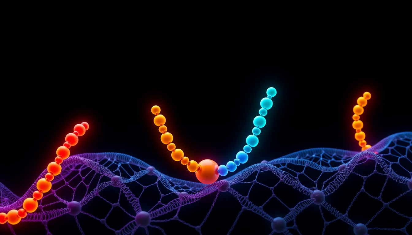
KPV is a tripeptide: lysine, proline, valine, in that order. It is the final three residues of α-melanocyte-stimulating hormone (α-MSH), a thirteen-residue hormone cut from the precursor protein pro-opiomelanocortin, and the research literature often writes it as α-MSH(11-13). The fragment's activity on its own was first reported in 1989 by Hiltz and Lipton at UT Southwestern in the FASEB Journal.1 It is also one of the four components of the KLOW research blend; our KLOW overview covers the other three. This article takes KPV on its own: what the molecule is, what the cell and animal work reports, and where the human evidence stops.
Research-use-only context. This article summarizes published third-party scientific literature — the large majority of it conducted in cultured cells or animal models. It is not medical advice, not a therapeutic or performance claim, and not a usage guide. American Peptides products are sold strictly for in vitro laboratory research and are not for human or veterinary use.
What KPV is
α-MSH has the sequence Ser-Tyr-Ser-Met-Glu-His-Phe-Arg-Trp-Gly-Lys-Pro-Val. The hormone's melanocortin-receptor pharmacophore — the His-Phe-Arg-Trp motif at positions 6 to 9 — is what engages the MC1 receptor on melanocytes and drives pigmentation. KPV is positions 11 to 13, and it contains none of that motif. A 2010 review by Brzoska and colleagues at the University of Münster makes the point directly: KPV lacks the entire sequence motif required for binding to any of the known melanocortin receptors, yet in their reading it retains most of the anti-inflammatory capacity of the full hormone while displaying no pigmentary action at all.2 The same review is candid that the exact signalling mechanism used by KPV is unknown.2
Chemically, KPV is about as simple as a bioactive peptide gets: roughly 342 daltons, basic and net positive at physiological pH because of the lysine side-chain amine, with a proline kink in the backbone and a small hydrophobic valine C-terminus. Several of the studies below also use a variant with D-valine in place of L-valine, written KP-D-V, and the antimicrobial literature works with a dimer in which two KPV units are joined through a cysteine–cysteine linker, written (CKPV)2 or, in its acetylated form, [Ac-CKPV]2.4,8
What cell and tissue studies report
Macrophages: NF-κB inhibition without receptor binding
The clearest single experiment on the receptor question is a 2001 Biochemical Pharmacology study from Mandrika, Muceniece and Wikberg at Uppsala, using the mouse macrophage-like line RAW 264.7.3 Cells were stimulated with bacterial lipopolysaccharide plus interferon-γ, and three peptides were compared: full-length α-MSH, the N-terminal fragment α-MSH(1-10), and the C-terminal fragment α-MSH(11-13), which is KPV. All three reduced nitric oxide production. All three reduced nuclear translocation of NF-κB, and KPV did so across the nanomolar-to-micromolar range.3 Radioligand binding showed MC1 receptor sites on the RAW cells; α-MSH and α-MSH(1-10) competed for them, and KPV did not, even at the highest concentration tested. α-MSH and the 1-10 fragment raised intracellular cyclic AMP, the second messenger downstream of MC1R; KPV did not. The authors concluded that melanocortin peptides act through two separable mechanisms, one dependent on MC1R and cAMP and one cAMP-independent that blocks NF-κB translocation, and that KPV uses only the second.3
Keratinocytes: no cyclic AMP, a calcium signal instead
A 2004 Journal of Investigative Dermatology study from Sheffield asked the same question in skin cells.4 Elliott and colleagues exposed the HaCaT keratinocyte line and normal human keratinocytes to α-MSH, KPV, KP-D-V and ACTH peptides and measured cyclic AMP: none of the peptides raised it in either cell type. What they did see was a rapid, acute rise in intracellular calcium in response to all of the peptides, but only when an adenosine agonist that suppresses the cAMP pathway was present. But when the group stably expressed MC1R in Chinese hamster ovary cells, both α-MSH and the KPV peptides raised intracellular calcium in those cells too.4 So the keratinocyte data agree with the macrophage data that KPV does not signal through cAMP, but they leave open whether KPV can engage MC1R in some contexts through a calcium-coupled route.
Intestinal epithelium: uptake through the PepT1 transporter
The most mechanistically specific KPV work is a 2008 Gastroenterology paper from Didier Merlin's group, then at Emory, which started from transport rather than receptors.5 PepT1 is a di- and tripeptide transporter normally expressed in the small intestine and induced in the colon during inflammatory bowel disease. In human intestinal epithelial lines (Caco2-BBE and HT29-Cl.19A) and in Jurkat T cells stimulated with pro-inflammatory cytokines, nanomolar KPV reduced activation of NF-κB and MAP-kinase signalling — measured by luciferase reporter, Western blot, real-time RT-PCR and ELISA — and reduced pro-inflammatory cytokine secretion. Competition experiments with a radiolabelled PepT1 substrate and uptake kinetics with tritiated KPV showed that KPV is carried into the cells by PepT1, and the authors concluded that KPV acts via PepT1 in both immune and epithelial cells.5 In this model the peptide enters the cell and acts inside it, which would explain why no surface receptor has been found.
Antimicrobial assays
A separate literature looks at KPV as an antimicrobial peptide. Cutuli, Catania and Lipton reported in a 2000 Journal of Leukocyte Biology paper that α-MSH and KPV inhibited colony formation by Staphylococcus aureus and reduced viability and germ-tube formation of the yeast Candida albicans, across a broad concentration range that included picomolar levels.7 Cyclic AMP rose in peptide-exposed yeast and an adenylyl cyclase inhibitor partly reversed the killing, which the authors read as a cAMP-dependent effect in the microbe. In the same paper the peptides did not reduce killing of either organism by human neutrophils; they enhanced it.7 A 2005 structural study from the same group examined the dimer [Ac-CKPV]2, built by inserting a Cys-Cys linker between two KPV units. The dimer was active against azole-resistant Candida species, and proton NMR with restrained dynamics calculations suggested an extended backbone with a β-turn-like element.8 The antimicrobial literature is thin: two organisms and essentially one laboratory.
What animal models report
The 1989 Hiltz and Lipton paper is the origin point. Mice were challenged with picryl chloride on the ear, which produces a measurable swelling; graded exposure to α-MSH(11-13) inhibited that swelling in a graded fashion, compared against saline and a corticosteroid control.1 It is a small, early study with a single endpoint.
In the Dalmasso 2008 paper, mice with colitis induced by dextran sodium sulfate (DSS) or by trinitrobenzene sulfonic acid (TNBS) were compared with and without KPV exposure; inflammation was assessed histologically and by pro-inflammatory cytokine mRNA expression in the colon. The exposed groups showed a lower incidence of colitis and reduced cytokine mRNA.5 The same year, Kannengiesser and colleagues at Münster published in Inflammatory Bowel Diseases using two models: DSS colitis and CD45RB(hi) T-cell transfer colitis. Readouts were body weight, colon histology, and myeloperoxidase (MPO) activity in colonic tissue, a marker of neutrophil infiltration. In DSS colitis the KPV-exposed mice recovered earlier and regained body weight more strongly; histological inflammatory infiltrates were reduced, and MPO activity was significantly lower. In transfer colitis the exposed group also recovered, regained weight and showed fewer inflammatory changes on histology.6 The key receptor experiment used mice carrying a non-functional MC1 receptor (MC1Re/e). KPV exposure during DSS colitis rescued every animal in that group from death, which led the authors to conclude that the effects were at least partially independent of MC1R signalling.6 Read alongside the Mandrika binding data: whatever KPV is doing in these models, a working MC1 receptor is not required.
Three caveats. These are chemically induced or cell-transfer models, not spontaneous disease. The readouts — histology scores, MPO, cytokine mRNA, body weight — are inflammation markers, not outcomes (our inflammation biomarkers explainer covers the distinction). And the two 2008 studies are most of the colitis evidence.
Where the human evidence stops
It stops before it starts. The reviewers closest to this compound write that the exact signalling mechanism used by KPV is currently unknown, and frame the tripeptides as candidates worth developing, not as agents with demonstrated effects in people.2 A PubMed search for KPV returns cell work, mouse work, peptide chemistry and delivery-matrix engineering, and no published controlled human study of KPV itself. The dimer [Ac-CKPV]2 was proposed as a candidate antifungal and studied structurally,8 but that is a different molecule, and its development status does not transfer to the monomer.
KPV is not approved by the FDA for any indication. It is an investigational research compound. Human data for α-MSH and its synthetic analogues concern different molecules carrying the pharmacophore KPV lacks, and do not transfer. Nothing in the cell or mouse literature above establishes what KPV does in a human being, because those studies have not been run.
What is not established
- What KPV binds. No receptor or intracellular target has been identified; the PepT1 data explain entry into the cell, not action once inside.2,5
- How KPV blocks NF-κB translocation once inside the cell, and whether the same mechanism operates across cell types.
- Whether the calcium response in MC1R-transfected cells means KPV can engage MC1R under some conditions, which would complicate the receptor-independent account.4
- Whether the monomer's antimicrobial activity is robust across assay conditions and across common chemical modifications such as N-terminal acetylation or C-terminal amidation.
- Any human pharmacology, safety or efficacy data at all.
Frequently Asked Questions
Is KPV the same as α-MSH?
No. α-MSH is a thirteen-residue hormone; KPV is its last three residues, Lys-Pro-Val. KPV lacks the His-Phe-Arg-Trp motif that α-MSH uses to bind melanocortin receptors, and in cell studies it does not compete for MC1R binding or raise cyclic AMP the way the full hormone does.
Does KPV work through melanocortin receptors?
The published evidence says largely not. In macrophages KPV did not bind MC1R sites or raise cAMP yet still blocked NF-κB translocation; in mice lacking a functional MC1 receptor KPV still altered the course of DSS colitis. Intestinal cell work points instead to uptake through the PepT1 transporter and an intracellular site of action; the actual target is unidentified.
What is PepT1 and why does it matter for KPV?
PepT1 is a transporter for di- and tripeptides expressed in the small intestine and induced in the colon during inflammation. Because KPV is a tripeptide, PepT1 carries it into epithelial and immune cells, which is the mechanism Merlin's group proposed for how it reaches the inside of a cell to act on NF-κB.
Is KPV approved by the FDA?
No. KPV is not approved by the FDA for any indication. There are no published controlled human studies of KPV; the evidence consists of cultured-cell, mouse and microbiology work. American Peptides supplies it strictly for in vitro research.
Citations
- Hiltz ME, Lipton JM. “Antiinflammatory activity of a COOH-terminal fragment of the neuropeptide alpha-MSH.” FASEB J. 1989;3(11):2282–2284. PubMed: PMID 2550304
- Brzoska T, Böhm M, Lügering A, Loser K, Luger TA. “Terminal signal: anti-inflammatory effects of α-melanocyte-stimulating hormone related peptides beyond the pharmacophore.” Adv Exp Med Biol. 2010;681:107–116. PubMed: PMID 21222263
- Mandrika I, Muceniece R, Wikberg JE. “Effects of melanocortin peptides on lipopolysaccharide/interferon-gamma-induced NF-kappaB DNA binding and nitric oxide production in macrophage-like RAW 264.7 cells: evidence for dual mechanisms of action.” Biochem Pharmacol. 2001;61(5):613–621. PubMed: PMID 11239505
- Elliott RJ, Szabo M, Wagner MJ, Kemp EH, MacNeil S, Haycock JW. “alpha-Melanocyte-stimulating hormone, MSH 11-13 KPV and adrenocorticotropic hormone signalling in human keratinocyte cells.” J Invest Dermatol. 2004;122(4):1010–1019. PubMed: PMID 15102092
- Dalmasso G, Charrier-Hisamuddin L, Nguyen HT, Yan Y, Sitaraman S, Merlin D. “PepT1-mediated tripeptide KPV uptake reduces intestinal inflammation.” Gastroenterology. 2008;134(1):166–178. PubMed: PMID 18061177
- Kannengiesser K, Maaser C, Heidemann J, et al. “Melanocortin-derived tripeptide KPV has anti-inflammatory potential in murine models of inflammatory bowel disease.” Inflamm Bowel Dis. 2008;14(3):324–331. PubMed: PMID 18092346
- Cutuli M, Cristiani S, Lipton JM, Catania A. “Antimicrobial effects of alpha-MSH peptides.” J Leukoc Biol. 2000;67(2):233–239. PubMed: PMID 10670585
- Catania A, Grieco P, Randazzo A, et al. “Three-dimensional structure of the alpha-MSH-derived candidacidal peptide [Ac-CKPV]2.” J Pept Res. 2005;66(1):19–26. PubMed: PMID 15946192
Related reading
- The KLOW Research Blend (GHK-Cu, BPC-157, TB-500, KPV): What the Component Studies Show
- Inflammation Biomarkers Explained: A Science Guide
- GHK-Cu (Copper Peptide) Research: What the Studies Actually Show
- How Signaling Peptides Work: A Complete Science Explainer
- BPC-157 Research: What the Published Studies Actually Show
This article is for laboratory research reference only. American Peptides products are sold strictly for in vitro research. Not for human consumption.
Related research
Compliance Notice: American Peptides products are sold strictly for laboratory and academic research purposes only. They are not intended for human or veterinary consumption, diagnosis, treatment, or prevention of any disease. All content on this page is educational in nature and does not constitute medical advice or product claims. Researchers are responsible for handling these compounds in accordance with their institution’s safety protocols and applicable laws.