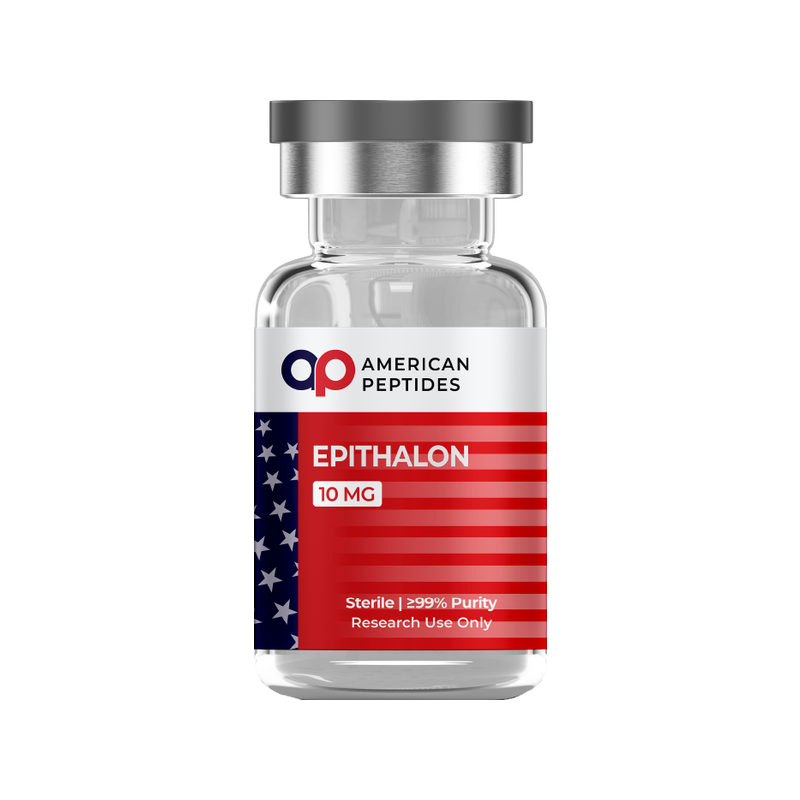


Epithalon — also spelled epitalon, and catalogued in the primary literature as the AEDG tetrapeptide Ala-Glu-Asp-Gly — is one of the oldest peptides still circulating in longevity discussion. Almost every claim attached to it online traces back to a single line of work: the St Petersburg Institute of Bioregulation and Gerontology, where the sequence was designed in the 1990s. This summary surveys what the peer-reviewed studies establish about how the molecule was built, what cultured-cell assays report about telomerase and gene expression, what animal models report about melatonin and the pineal gland, and where the evidence stops. For the evidence-first format used across this series, see our MOTS-c research review.
Research-use-only context. This article summarizes published third-party scientific literature — the large majority of it conducted in cultured cells or animal models. It is not medical advice, not a therapeutic or performance claim, and not a usage guide. American Peptides products are sold strictly for in vitro laboratory research and are not for human or veterinary use.
What epithalon is
Epithalon is a synthetic peptide of four residues: alanine, glutamic acid, aspartic acid and glycine. It was not isolated from tissue. According to Khavinson’s monograph-length 2002 review in Neuro Endocrinology Letters, the sequence was designed from the amino-acid composition of epithalamin, a polypeptide extract of bovine pineal gland the institute had studied for decades.2 The group built a family of short peptides from the compositions of tissue extracts — dipeptides for the thymus, tetrapeptides for heart, liver, cerebral cortex and pineal gland — and reported that each stimulated the in vitro outgrowth of explants from its own source organ. The same Ala-Glu-Asp-Gly sequence emerged for both the retina and the pineal gland, which the authors attribute to the shared embryonic origin of the two tissues.2 A 2025 review from the Medical University of Warsaw adds that the tetrapeptide was synthesized from epithalamin’s composition before the same sequence was identified within the pineal polypeptide complex itself.1
Epithalon is not a hormone and has no defined cell-surface receptor. The Warsaw review, surveying twenty-five years of in vitro, in vivo and in silico work, lists the activities that have been reported — a direct influence on melatonin synthesis, altered interleukin-2 mRNA levels, modulation of the mitogenic activity of murine thymocytes, and increased activity of several enzymes including acetylcholinesterase, butyrylcholinesterase and telomerase — and then states plainly that it “remains uncertain whether these are the sole mechanisms of action of this compound.” The same authors note that physico-chemical and structural investigations of the peptide remain “quite limited” relative to the volume of biological reports.1 For context, see why peptides are studied in aging research.
What the research reports
Molecular design and the proposed mechanism
The St Petersburg group’s explanation for how a tetrapeptide could affect tissue function is transcriptional. Khavinson’s 2002 review sets out a “peptide theory of ageing” in which aging is described as an evolutionarily determined shift in gene expression that impairs the synthesis of regulatory and tissue-specific peptides, and proposes that short peptides act on transcriptional machinery shared by the pineal gland and the retina.2 This is a hypothesis built on observed changes in gene expression, not a demonstrated receptor pathway, and the Warsaw reviewers classify the reported effects as arising from “both specific and nonspecific mechanisms.”1
Telomerase and gene-expression studies in cultured cells
The telomerase literature is what made epithalon famous, and it began with two short reports in the Bulletin of Experimental Biology and Medicine. In 2003, Khavinson, Bondarev and Butyugov reported that adding the peptide to a telomerase-negative human fetal fibroblast culture induced expression of the enzyme’s catalytic subunit, measurable telomerase activity, and telomere elongation.3 A 2004 follow-up from the same authors used primary pulmonary fibroblasts from a 24-week fetus, which lost proliferative capacity at the 34th passage with telomeres appreciably shorter than at passage 10. Adding epithalon to the aging cultures was reported to lengthen telomeres to sizes comparable to early passages, and the exposed cells completed ten additional divisions, reaching 44 passages and continuing to divide. The authors framed this as overcoming the Hayflick limit.4 Both papers are brief, come from one laboratory, and their abstracts give no replicate numbers; for two decades the finding sat without independent confirmation.
That changed in 2025. A group at Brunel University London, with no connection to the originating institute, published a quantitative study in Biogerontology using the breast cancer lines 21NT and BT474 alongside normal epithelial and fibroblast cells. By qPCR and immunofluorescence they reported concentration-dependent telomere lengthening in the normal cells, accompanied by upregulation of hTERT mRNA and telomerase enzyme activity. In the cancer lines, telomeres also lengthened — but through activation of the alternative lengthening of telomeres (ALT) pathway rather than telomerase, with only a minor ALT signal in normal cells.5 This is the first independent replication of the core claim, and it cuts both ways: telomere lengthening in cancer cells by a telomerase-independent route is not an effect anyone should call uniformly desirable. Telomere maintenance is one of the double-edged hallmarks discussed in the biology of aging and longevity.
Melatonin, pineal and circadian findings in animal models
Because the peptide was modeled on a pineal extract, the earliest animal work asked whether it affected the pineal gland’s output. The most-cited study is a 2001 report by Khavinson, Goncharova and Lapin in female rhesus macaques (Macaca mulatta) of different ages. Measuring melatonin and cortisol by immunoassay, the authors reported that epithalon significantly stimulated evening melatonin synthesis in the senescent animals, and that the circadian rhythm of cortisol secretion, which had become disorganized with age, moved back toward the pattern seen in younger monkeys.6 Khavinson’s 2002 review summarizes the same work as a restoration of the circadian rhythms of melatonin and cortisol in old rhesus monkeys, and lists increased melatonin production as the first effect of the parent extract epithalamin in rats.2 The Warsaw review likewise lists a direct influence on melatonin synthesis among the peptide’s best-supported activities.1
The limitation is familiar: a single laboratory, a primate study whose abstract does not report group sizes, and a readout that varies considerably between individuals. Melatonin rhythm is a measurable marker of pineal function (see the biology of sleep and recovery), but a shift in a hormone rhythm in aged macaques is a physiological observation, not an outcome.
Other animal-model findings
The survival study most often quoted is Anisimov and colleagues’ 2003 paper in Biogerontology, and its results are more modest than the summaries of it. Female outbred Swiss-derived SHR mice, 54 per group, were exposed to epithalon or saline from three months of age until natural death. Epithalon did not change food consumption, body weight or mean life span. What the authors did report was a slower age-related loss of estrous function; a 17.1% reduction in the frequency of chromosome aberrations in bone-marrow cells; a 13.3% longer life span among the last 10% of survivors; and a 12.3% higher maximum life span than controls. Total spontaneous tumor incidence was unchanged, though leukemia developed 6.0-fold less often in the peptide group.7 In other words, the headline “lifespan” result rests on the tail of a survival curve in one sex of one mouse strain, from one laboratory, with the mean unchanged. Khavinson’s review groups this with fruit-fly data under the group’s own term, “geroprotector” activity2 — a label that belongs to the authors, not the field.
A second independent group has since examined the peptide in a different model. In 2022, Yue and colleagues at Shanxi Medical University and the Chinese Academy of Sciences, publishing in Aging, cultured mouse oocytes through post-ovulatory aging in vitro with and without epithalon in the medium. They reported lower intracellular reactive oxygen species, fewer spindle defects and abnormal cortical-granule distributions at 12 and 24 hours, higher mitochondrial membrane potential and mitochondrial DNA copy number, and less apoptosis by 24 hours, and attributed the effect to modulation of mitochondrial activity and ROS levels.8 It is an in vitro oocyte model, not an animal outcome, but it adds a mitochondrial readout the original St Petersburg work never measured (see what mitochondrial health means).
The human-evidence gap
Here candor matters more than enthusiasm. The large majority of the epithalon literature — the telomerase reports, the macaque study, the mouse survival study, the mechanistic theory — comes from one research group with overlapping authors, much of it published as brief communications whose abstracts omit sample sizes, statistics and methods. Khavinson’s own 2002 review states that clinical observations exist for the parent extract epithalamin and for epithalon in degenerative retinal conditions.2 Those observations were generated by the same institute, were not independent randomized trials in the sense a regulator would recognize, and cannot be evaluated from the published abstracts. Independent replication arrived only in 2022 and 2025, and both studies are in vitro.5,8 The Warsaw reviewers, otherwise favorable, close by noting that the mechanism remains uncertain and structural data are thin.1
No regulator has approved epithalon for any purpose. It is not FDA-approved, has no marketing authorization in the European Union, and appears in no pharmacopoeia. That matters for how research-grade material should be understood: a vial of synthetic Ala-Glu-Asp-Gly supplied to a laboratory has no human data of its own, because none of the studies above was conducted with it, and none generated human safety or outcome data that could transfer to it. The animal findings summarized here describe hormone rhythms in aged macaques and the tail of a survival curve in one mouse strain. They do not describe what happens in a person; the gap between a longer-lived cohort of mice and a healthier human life is the subject of healthspan versus lifespan, not something a tetrapeptide study can resolve.
The takeaway
Epithalon is a well-defined molecule with a clear origin story: a four-residue sequence designed from the composition of a bovine pineal extract, proposed to act through gene transcription. Its strongest laboratory finding — telomerase upregulation and telomere lengthening in cultured human cells — was reported by one group in 2003 and 2004 and independently confirmed, with nuances about ALT activity in cancer lines, in 2025. Its animal literature reports altered evening melatonin and cortisol rhythm in aged macaques and a shift in maximum, but not mean, life span in one mouse strain. What it lacks is independent, controlled human evidence of any kind, and regulatory approval anywhere. American Peptides supplies epithalon strictly for in vitro research; sequence, formula and handling specifications are on the epithalon reference monograph.
Frequently Asked Questions
What is the amino-acid sequence of epithalon?
Ala-Glu-Asp-Gly, abbreviated AEDG: alanine, glutamic acid, aspartic acid and glycine. It is a synthetic tetrapeptide designed from the amino-acid composition of epithalamin, a bovine pineal extract.
Does epithalon activate telomerase?
In cultured cells, according to the available reports. The originating group described induction of the telomerase catalytic subunit, enzyme activity and telomere elongation in human fetal fibroblasts in 2003 and 2004; an independent 2025 Brunel University study reported hTERT upregulation and telomere lengthening in normal human cells, with ALT-pathway activation in breast cancer lines. All of this is in vitro.
Is epithalon the same as epithalamin?
No. Epithalamin is a multi-component polypeptide extract of bovine pineal gland. Epithalon is a single synthetic tetrapeptide whose sequence was modeled on the amino-acid composition of that extract. The two literatures overlap because the same group studied both.
Is epithalon approved by the FDA?
No. It is not FDA-approved and holds no regulatory approval for any use in any jurisdiction. American Peptides supplies it strictly for in vitro research.
Citations
- Araj SK, et al. “Overview of Epitalon-Highly Bioactive Pineal Tetrapeptide with Promising Properties.” Int J Mol Sci. 2025;26(6):2691. PubMed: PMID 40141333
- Khavinson VKh. “Peptides and Ageing.” Neuro Endocrinol Lett. 2002;23 Suppl 3:11–144. PubMed: PMID 12374906
- Khavinson VKh, et al. “Epithalon peptide induces telomerase activity and telomere elongation in human somatic cells.” Bull Exp Biol Med. 2003;135(6):590–592. PubMed: PMID 12937682
- Khavinson VKh, et al. “Peptide promotes overcoming of the division limit in human somatic cell.” Bull Exp Biol Med. 2004;137(5):503–506. PubMed: PMID 15455129
- Al-Dulaimi S, et al. “Epitalon increases telomere length in human cell lines through telomerase upregulation or ALT activity.” Biogerontology. 2025;26(5):178. PubMed: PMID 40908429
- Khavinson V, et al. “Synthetic tetrapeptide epitalon restores disturbed neuroendocrine regulation in senescent monkeys.” Neuro Endocrinol Lett. 2001;22(4):251–254. PubMed: PMID 11524632
- Anisimov VN, et al. “Effect of Epitalon on biomarkers of aging, life span and spontaneous tumor incidence in female Swiss-derived SHR mice.” Biogerontology. 2003;4(4):193–202. PubMed: PMID 14501183
- Yue X, et al. “Epitalon protects against post-ovulatory aging-related damage of mouse oocytes in vitro.” Aging (Albany NY). 2022;14(7):3191–3202. PubMed: PMID 35413689
Related reading
- The Biology of Aging and Longevity
- Why Peptides Are Studied in Aging Research
- The Biology of Sleep and Recovery
This article is for laboratory research reference only. American Peptides products are sold strictly for in vitro research. Not for human consumption.
Related research
Compliance Notice: American Peptides products are sold strictly for laboratory and academic research purposes only. They are not intended for human or veterinary consumption, diagnosis, treatment, or prevention of any disease. All content on this page is educational in nature and does not constitute medical advice or product claims. Researchers are responsible for handling these compounds in accordance with their institution’s safety protocols and applicable laws.