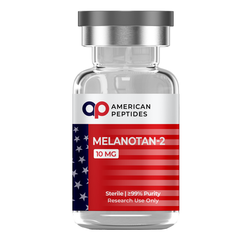

Melanotan II (MT-II) is a synthetic cyclic heptapeptide, Ac-Nle-Asp-His-D-Phe-Arg-Trp-Lys-NH2, built from residues 4–10 of the thirteen-residue pituitary hormone alpha-melanocyte-stimulating hormone (α-MSH) and closed into a ring by a lactam bond between its aspartate and lysine side chains. It was designed in 1989 at the University of Arizona by Victor Hruby’s chemistry group with the physiologist Mac Hadley, building on Tomi Sawyer’s 1980 linear analogue [Nle4, D-Phe7]-α-MSH. Pharmacologically it is a non-selective agonist at four of the five melanocortin receptors: MC1R, MC3R, MC4R and MC5R. This article covers the chemistry that produced it, what receptor and pigment-cell assays report, what rodent studies measured, and where the human evidence stops. For how peptide ligands engage G-protein-coupled receptors, see our receptor pathways primer.
Research-use-only context. This article summarizes published third-party scientific literature — the large majority of it conducted in cultured cells or animal models. It is not medical advice, not a therapeutic or performance claim, and not a usage guide. American Peptides products are sold strictly for in vitro laboratory research and are not for human or veterinary use.
What Melanotan II is
The parent hormone, α-MSH (Ac-Ser-Tyr-Ser-Met-Glu-His-Phe-Arg-Trp-Gly-Lys-Pro-Val-NH2), is cleaved from pro-opiomelanocortin, and its His-Phe-Arg-Trp core at positions 6–9 is the segment every melanocortin receptor recognises. Its methionine-4 oxidises readily and serum enzymes degrade it quickly, so its activity in bioassays is brief.
The first step toward MT-II was an observation. In a 1980 PNAS paper, Sawyer, Hruby, Hadley and colleagues reported that heat-alkali exposure prolonged the activity of α-MSH on frog skin, and that gas chromatography of the hydrolysed peptide showed partial racemisation at positions 4 and 7. Replacing methionine-4 with the oxidation-resistant isostere norleucine and L-phenylalanine-7 with its D-enantiomer gave [Nle4, D-Phe7]-α-MSH, which reproduced the prolonged activity, resisted serum enzymes, and was 26 times as potent as α-MSH in a mouse melanoma adenylate cyclase assay.1 That analogue, NDP-MSH (later Melanotan I), is MT-II’s direct ancestor.
In a 1989 Journal of Medicinal Chemistry paper, Al-Obeidi, Castrucci, Hadley and Hruby used structure–activity data and molecular dynamics simulations of NDP-MSH and a disulfide-bridged [Cys4, Cys10] analogue to design cyclic lactam fragments spanning residues 4–10 or 4–13. The ring joined a Glu or Asp side chain at position 5 to a Lys, Orn, Dab or Dpr side chain at position 10, with Nle4 and D-Phe7 retained. Of seven cyclic peptides, the aspartate–lysine bridge, Ac-[Nle4, Asp5, D-Phe7, Lys10]-α-MSH(4–10)-NH2 — the compound now called MT-II — was about 90 times as potent as α-MSH on lizard skin. Ring size mattered: the 23-membered ring was optimal, shrinking or enlarging it cut potency, and only the 23- and 24-membered rings showed prolonged residual activity after washout.2 The logic is conformational: a lactam locks the His-Phe-Arg-Trp core into the turn the receptor prefers, and unlike a disulfide it cannot be opened by reduction.
The receptor family was cloned after the compound was made. As Hruby and Cone’s groups summarised in 1995, MC2R recognises only ACTH, while MC1R, MC3R, MC4R and MC5R all respond to α-MSH, NDP-MSH and the cyclic lactam.3 MT-II was optimised on pigment cells for potency and stability, not for subtype selectivity, and it activates all four.
What cell and tissue studies report
Receptor assays in transfected cells
Much of the receptor literature uses MT-II as the scaffold from which selective ligands were carved. The 1995 Hruby paper replaced D-Phe7 with bulkier aromatic D-amino acids and tested the products on frog skin and on four cloned mammalian receptors expressed in cells. The D-2′-naphthylalanine-7 analogue, later known as SHU9119, was a potent antagonist at MC4R (pA2 9.3) and a weaker one at MC3R (pA2 8.3), yet remained a full agonist at MC1R and MC5R. The D-p-iodophenylalanine-7 analogue was a potent agonist at human MC1R (EC50 0.055 nM) while antagonising human MC4R and MC3R; the D-p-chloro and D-p-fluoro versions were full agonists everywhere.3 Position 7 is thus a switch between agonism and subtype-selective antagonism.
A 1999 Biochemical and Biophysical Research Communications paper from Merck (Bednarek and colleagues) examined the other end of the molecule in binding and activation assays on human MC3R, MC4R and MC5R. Replacing Nle4 with proline gave [Pro4]MTII, which kept MT-II’s affinity and potency at MC4R but lost about 400-fold at MC5R and about 20-fold at MC3R. Other N-terminal substitutions gave the same pattern, and the authors concluded that the N-terminal fragment outside the lactam ring has virtually no effect on MC4R but is essential for full potency at MC5R.4 Together the two papers give a working map: the ring carries MC4R recognition, position 7 governs agonism versus antagonism at MC3R and MC4R, and the exocyclic tail tunes MC5R.
Melanin synthesis in cultured melanocytes
The pathway downstream of MC1R is the best characterised: the receptor couples to Gs, cyclic AMP rises, and cAMP increases the expression and activity of tyrosinase, the rate-limiting enzyme of melanin synthesis. The 1980 Sawyer paper reported both readouts for the linear Nle4, D-Phe7 analogue in mouse melanoma cells: adenylate cyclase activation and tyrosinase stimulation.1
Human melanocytes proved harder. A 1994 Journal of Cell Science study by Hunt, Todd, Cresswell and Thody cultured human melanocytes without the artificial mitogens of standard media and incubated them with α-MSH or NDP-MSH over three days; morphology shifted from mainly bipolar to dendritic in roughly two-thirds of cultures within 18 hours. NDP-MSH produced concentration-related increases in tyrosinase activity and melanin content, with tyrosinase the more responsive measure, and the two peptides appeared roughly equipotent on human cells. Not every culture responded, responsiveness was unrelated to basal tyrosinase or melanin, and in some cultures the morphological and melanogenic responses were independent of one another.5 Note that this study used the linear analogue, not the cyclic one.
What animal models report
Amphibian and reptile skin bioassays
Before the receptors were cloned, potency was measured on living skin. In the frog (Rana pipiens) and lizard (Anolis carolinensis) bioassays, isolated skin darkens reversibly as melanosomes disperse within dermal melanophores, and the darkening is quantified by reflectance. Sawyer used frog skin to show reversible darkening with greatly prolonged activity for the Nle4, D-Phe7 analogue.1 Al-Obeidi ran the seven cyclic lactams on both species: the best were 90 to 100 times as potent as α-MSH on lizard skin but much less potent on frog skin, which the authors read as selectivity for the lizard pigment-cell receptor over the frog one.2 Hruby then used frog skin as a functional MC1R assay to define antagonism, reporting pA2 values above 10 for the bulky-aromatic position-7 analogues.3
Mouse studies
The rodent literature on MT-II is mostly not about pigment. In a 1997 Nature paper, Fan, Boston, Kesterson, Hruby and Cone asked why dominant agouti alleles produce a metabolic syndrome in mice. The agouti peptide antagonises MC1R in skin, switching melanocytes away from eumelanin synthesis, and also the hypothalamic MC4R. Using MT-II and SHU9119 as an agonist–antagonist pair, the group measured food intake in four mouse models with elevated intake: fasted C57BL/6J, ob/ob, agouti Ay, and neuropeptide Y-stimulated mice. In every model, intake fell in the groups exposed to MT-II; with SHU9119 present the reduction was completely blocked; and SHU9119 alone increased feeding after a fast. The authors concluded that melanocortinergic neurons exert a tonic inhibitory signal on feeding behaviour.6 The methodological lesson: because MT-II activates four subtypes, attribution to the central receptors rested on the antagonist control and the agouti genetics, not on the agonist alone.
Where the human evidence stops
Reviewers in dermatology have been direct. A 2017 review in the International Journal of Dermatology by Habbema, Halk, Neumann and Bergman describes the synthetic α-MSH analogues sold as melanotan I and II as unregulated and untested, and notes open questions about the preparation and identity of products in circulation.8
The controlled human data on MT-II itself come down to one pilot. In a 1996 Life Sciences paper, Dorr and colleagues reported a single-blind, placebo-controlled phase I pilot in three healthy male volunteers. Two of the three showed increased skin pigmentation, measured by quantitative reflectance and visual assessment, one week after the exposure period ended. Adverse findings reported included somnolence and fatigue in one volunteer at the highest exposure level, mild nausea at most levels, and a stretching-and-yawning complex.7 No larger controlled trial of MT-II for any pigment-cell endpoint has been published.
MT-II is not approved by the FDA for any purpose. It is an investigational compound, and products sold outside regulated channels have been the subject of safety warnings from multiple national health regulators. The Habbema review collects the case literature behind those warnings: melanocytic changes in existing moles and newly emerging dysplastic naevi associated with unregulated melanotan I and II, and four case reports of melanoma arising from existing moles, while stressing that conclusive evidence of a causal link is lacking.8 Those observations concern products of unknown composition, not a characterised laboratory reagent. Our guide to spotting a grey-market peptide vendor covers why composition matters.
What is not established
- Whether the cultured human melanocyte findings obtained with linear NDP-MSH are reproduced quantitatively by the cyclic lactam; the two have not been compared side by side in primary human melanocytes.
- Why a subset of human melanocyte cultures does not respond to MSH peptides, and whether MC1R genotype accounts for it.
- Which receptor subtype mediates each rodent observation made with MT-II, since attribution depends on antagonist and knockout controls.
- Whether the melanocytic changes in case reports reflect receptor pharmacology, impurities in unregulated products, or coincidence.
- Any controlled human outcome beyond a three-person pilot.
Frequently Asked Questions
What is the chemical difference between Melanotan I and Melanotan II?
Melanotan I (NDP-MSH, afamelanotide) is the full thirteen-residue linear α-MSH sequence with norleucine at position 4 and D-phenylalanine at position 7. Melanotan II is a seven-residue fragment (positions 4–10) carrying the same two substitutions, cyclised through a lactam bond between aspartate-5 and lysine-10 to form a 23-membered ring.
Which receptors does Melanotan II act on?
In cloned-receptor assays it is an agonist at MC1R, MC3R, MC4R and MC5R with little selectivity between them. MC2R, the adrenocorticotropin receptor, does not respond to it. Selective ligands such as SHU9119 and [Pro4]MTII were made by modifying the MT-II scaffold at position 7 or at the N-terminus.
What do pigment-cell studies measure?
In cultured melanocytes, the standard readouts are cyclic AMP, tyrosinase activity, melanin content and cell morphology. In frog and lizard skin bioassays, the readout is reversible skin darkening caused by melanosome dispersion within melanophores, quantified by reflectance.
Is Melanotan II approved by the FDA?
No. Melanotan II is not approved by the FDA for any purpose. It is an investigational compound, and unregulated products have been the subject of safety warnings from multiple national health regulators. American Peptides supplies it strictly for in vitro research.
Citations
- Sawyer TK, Sanfilippo PJ, Hruby VJ, et al. (1980). “4-Norleucine, 7-D-phenylalanine-alpha-melanocyte-stimulating hormone: a highly potent alpha-melanotropin with ultralong biological activity.” Proc Natl Acad Sci U S A. 77(10):5754–5758. PubMed: PMID 6777774
- Al-Obeidi F, Castrucci AM, Hadley ME, Hruby VJ. (1989). “Potent and prolonged acting cyclic lactam analogues of alpha-melanotropin: design based on molecular dynamics.” J Med Chem. 32(12):2555–2561. PubMed: PMID 2555512
- Hruby VJ, Lu D, Sharma SD, et al. (1995). “Cyclic lactam alpha-melanotropin analogues of Ac-Nle4-cyclo[Asp5, D-Phe7,Lys10] alpha-melanocyte-stimulating hormone-(4-10)-NH2 with bulky aromatic amino acids at position 7 show high antagonist potency and selectivity at specific melanocortin receptors.” J Med Chem. 38(18):3454–3461. PubMed: PMID 7658432
- Bednarek MA, Macneil T, Kalyani RN, et al. (1999). “Analogs of MTII, lactam derivatives of alpha-melanotropin, modified at the N-terminus, and their selectivity at human melanocortin receptors 3, 4, and 5.” Biochem Biophys Res Commun. 261(1):209–213. PubMed: PMID 10405347
- Hunt G, Todd C, Cresswell JE, Thody AJ. (1994). “Alpha-melanocyte stimulating hormone and its analogue Nle4DPhe7 alpha-MSH affect morphology, tyrosinase activity and melanogenesis in cultured human melanocytes.” J Cell Sci. 107(Pt 1):205–211. PubMed: PMID 8175909
- Fan W, Boston BA, Kesterson RA, Hruby VJ, Cone RD. (1997). “Role of melanocortinergic neurons in feeding and the agouti obesity syndrome.” Nature. 385(6612):165–168. PubMed: PMID 8990120
- Dorr RT, Lines R, Levine N, et al. (1996). “Evaluation of melanotan-II, a superpotent cyclic melanotropic peptide in a pilot phase-I clinical study.” Life Sci. 58(20):1777–1784. PubMed: PMID 8637402
- Habbema L, Halk AB, Neumann M, Bergman W. (2017). “Risks of unregulated use of alpha-melanocyte-stimulating hormone analogues: a review.” Int J Dermatol. 56(10):975–980. PubMed: PMID 28266027
Related reading
- What Is a Peptide? A Researcher’s Primer on Structure and Signaling
- Receptor Pathways: A Researcher’s Primer
- Amino Acid Signaling and Receptor Biology: A Research Primer
- How to Spot a Grey-Market Peptide Vendor: 9 Red Flags Researchers Should Know
This article is for laboratory research reference only. American Peptides products are sold strictly for in vitro research. Not for human consumption.
Related research
Compliance Notice: American Peptides products are sold strictly for laboratory and academic research purposes only. They are not intended for human or veterinary consumption, diagnosis, treatment, or prevention of any disease. All content on this page is educational in nature and does not constitute medical advice or product claims. Researchers are responsible for handling these compounds in accordance with their institution’s safety protocols and applicable laws.