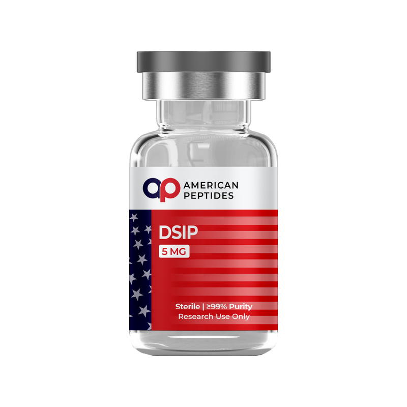

DSIP — delta sleep-inducing peptide — has one of the strangest biographies in neuropeptide research. Its nine-residue sequence has been known since 1977 and it has been studied in rabbits, rats, mice, cats and guinea pigs, yet no gene that encodes it has ever been found, no receptor has been identified, and the property it was named for has proved difficult to reproduce. Most online writing about DSIP skips those problems. This summary surveys what the peer-reviewed literature establishes about how the molecule was found, where DSIP-like material appears in the body, what animal electrophysiology and biochemistry report, and where the evidence stops. For the evidence-first format used across this series, see our Selank research review.
Research-use-only context. This article summarizes published third-party scientific literature — the large majority of it conducted in cultured cells or animal models. It is not medical advice, not a therapeutic or performance claim, and not a usage guide. American Peptides products are sold strictly for in vitro laboratory research and are not for human or veterinary use.
What DSIP is
DSIP is a nonapeptide with the sequence Trp-Ala-Gly-Gly-Asp-Ala-Ser-Gly-Glu and a molecular weight of about 849.1,3 Its discovery grew out of a long programme of “humoral transmission of sleep” experiments in Basel. Between 1963 and 1970, Marcel Monnier’s group established that blood drawn from the cerebral venous sinus of rabbits held in slow-wave sleep by electrical stimulation of the thalamus, then dialysed, carried a factor that increased delta-band EEG activity in recipient rabbits.2 Between 1970 and 1977 the group isolated, characterised and synthesised the nonapeptide responsible for that effect.2
The 1977 paper by Guido Schoenenberger and Monnier in Proceedings of the National Academy of Sciences reported the sequence, synthesised the peptide along with five possible metabolic fragments (residues 1–8, 2–9, 2–8, 1–4 and 5–9), two nonapeptide analogues with two amino acids exchanged, and a related tripeptide (Trp-Ser-Glu), and tested all nine synthetic peptides in rabbits under double-blind conditions. Fifty-eight rabbits including controls were evaluated, with neocortical and archicortical EEG analysed by fast Fourier transform. Only the synthetic nonapeptide itself produced significant, specific enhancement of delta and spindle EEG patterns, which is how it earned its name.1
What was never found is the machinery around it. Kovalzon and Strekalova’s 2006 review in the Journal of Neurochemistry states the problem plainly: the DSIP gene, a precursor protein and a receptor have never been isolated; the structure “is different from any other known representative of the various peptide families”; and the link between DSIP and sleep “has never been further characterized.”4 That unresolved receptor question frames everything that follows.
What the research reports
Molecular structure and stability
In Schoenenberger’s 1984 account of the Basel programme, analogues with exchanged residues, peptides shortened by one or two amino acids, and breakdown products all weakened or abolished the EEG effect in animals, “suggesting a close structure-specificity.”2 The same review describes a phosphorylated derivative, DSIP-P, in which the serine at position 7 carries a phosphate group; the group reported it as many-fold more potent than the parent peptide, and detected both DSIP and DSIP-P in human cerebrospinal fluid.2
Stability is a genuine issue for a short, unprotected peptide. In brain slices and homogenates, the half-life for proteolytic removal of the N-terminal tryptophan was about 15 minutes.2 Endogenous DSIP-like immunoreactive material in plasma, urine and CSF, by contrast, was found bound to a larger protein, which the authors interpreted as a carrier that shields the peptide from proteolysis.2 The tension between a fragile free peptide and protected endogenous material runs through the field; Graf and Kastin’s 1984 review in Neuroscience & Biobehavioral Reviews accordingly describes most in vivo measurements as “DSIP-like material” detected by radioimmunoassay rather than confirmed free nonapeptide.3
Where the peptide is found, and the receptor question
Immunohistochemistry has mapped DSIP-like immunoreactivity in several species. In the rat diencephalon, Skagerberg and colleagues found varicose nerve fibres carrying DSIP-like immunoreactivity throughout the mediobasal hypothalamus, most densely in the arcuate nucleus, the adjoining median eminence and the pituitary stalk. Even after colchicine pre-exposure, they could find no immunoreactive cell bodies at all, only fibres.5 The authors read this innervation pattern as a basis for a role in neuroendocrine regulation at the hypothalamic level, not specifically in sleep.5 Graf and Kastin add that DSIP-like material has been found in peripheral rat organs and in the plasma of several mammals.3
That anatomy makes the receptor question awkward. Kovalzon and Strekalova point out that the Geneva group’s histochemical studies place DSIP-like immunoreactivity in neurosecretory hypothalamic nuclei “that are not particularly relevant for sleep regulation.”4 Their proposed resolution is that an as-yet-unidentified DSIP-like peptide, rather than DSIP itself, accounts for both the immunoreactivity and the biological activity. They note that in their own earlier rabbit and rat work certain artificial structural analogues promoted slow-wave sleep while the native nonapeptide did not, and that a naturally occurring dermorphin-related decapeptide sharing five of nine positions with DSIP promoted slow-wave sleep in rabbits while its optical isomer suppressed it.4
What has been characterised is a transport mechanism rather than a receptor. In the vascularly perfused guinea pig forebrain, Zlokovic and colleagues measured unidirectional transfer of radiolabelled DSIP across the blood-brain barrier, with regional transfer constants of 0.93, 1.33 and 1.66 microlitres per minute per gram for parietal cortex, caudate nucleus and hippocampus. Adding unlabelled DSIP cut uptake to values comparable with mannitol, a marker that does not cross the barrier, and L-tryptophan, the peptide’s N-terminal residue, also inhibited transfer. The authors concluded that a high-affinity, saturable transport system carries DSIP across the barrier, with binding at specific sites on the brain capillary membrane.6 A saturable carrier implies that some protein recognises the peptide; it does not identify a signalling receptor.
Electrophysiology and sleep architecture in animal models
Beyond the founding 1977 rabbit EEG result,1 the two 1984 reviews extend the animal picture in three ways. First, the effect was species-dependent: rabbits, rats and mice showed mainly delta-sleep changes, whereas in cats the effect on REM sleep was more pronounced.2,3 Second, the response did not scale monotonically. Both reviews describe a U-shaped or parabolic response curve with distinct optima, which Schoenenberger contrasts with conventional pharmacological agents.2,3 Third, in animals DSIP also shifted the circadian rhythmicity of locomotor activity, brain transmitter concentrations, and plasma protein and cortisol levels.2
Graf and Kastin also list reported animal effects on brain neurotransmitter levels, hormone levels, and the activity and withdrawal of neuropharmacological agents,3 and Schoenenberger adds induction of monoamine oxidase A and RNA synthesis in brain, a rise in brain DSIP concentration during advanced hibernation, a fall in alcohol-dependent rats, and a specific electrophysiological effect on isolated rat and rabbit neurons.2 These observations underpin the peptide’s reputation. They are rabbit, rodent and cat EEG and biochemistry from the 1970s and 1980s, and the 2006 review regards the sleep-factor hypothesis built on them as “extremely poorly documented and still weak.”4
Stress-axis, mitochondrial and other animal-model findings
A second strand of the literature concerns stress physiology. In rats pre-exposed to chlorpromazine, morphine and pentobarbital, Graf, Kastin, Coy and Fischman found that DSIP significantly reduced the corticosterone release stimulated by corticotropin-releasing factor, but had no effect on corticosterone release stimulated by adrenocorticotropic hormone itself. They interpreted this as attenuation of CRF action at the level of the pituitary rather than the adrenal gland.7 It remains the cleanest mechanistic data point behind the claim that DSIP interacts with the stress axis.
Later Russian work moved to the mitochondrion. Khvatova and colleagues measured oxygen consumption polarographically in isolated rat brain mitochondria and found that DSIP significantly increased the rate of phosphorylating (state 3) respiration while leaving uncoupled respiration unchanged, raising the respiratory control ratio and the rate of ADP phosphorylation; the same pattern appeared in rat brain homogenates. In rats subjected to hypoxia, which on its own reduced state 3 respiration and ADP phosphorylation, prior exposure to DSIP prevented that reduction.8 The authors offer this as a possible mechanism behind stress-protective effects reported in rodents; it is an isolated-organelle and rodent finding, nothing more.8
The human-evidence gap
The animal literature above is real, but it is old, thin in places, and internally contested. Three facts matter most. First, no receptor, gene or precursor protein for DSIP has ever been identified, so there is no molecular target against which to test specificity.4 Second, replication is inconsistent: Kovalzon and Strekalova report that native DSIP failed to promote slow-wave sleep in their own rabbit and rat experiments while synthetic analogues succeeded.4 Third, the bulk of the primary literature dates from 1977 to the early 1990s, and later rodent work comes from a small number of laboratories, some of it on a formulated product based on DSIP rather than the peptide alone.2,3,8
Human observations exist but are sparse. Graf and Kastin’s 1984 review lists humans among the species in which delta-sleep changes were reported, and Schoenenberger’s group detected DSIP-like immunoreactive material in human plasma, urine, CSF and milk.2,3 Those were early reports, and the 2006 review’s judgement is that the link between DSIP and sleep “has never been further characterized.”4 There are no controlled human outcome data for any of the uses discussed on wellness forums, and DSIP is not approved by the FDA for any purpose.
Research-grade DSIP has no human data of its own. It is a reference compound for the questions the literature leaves open: what the saturable blood-brain-barrier carrier recognises, what the hypothalamic immunoreactivity is, and whether the corticosterone and mitochondrial findings reproduce in independent hands.
The takeaway
DSIP is a well-defined molecule with a poorly defined biology. Its sequence and structure–activity profile were settled by the Basel work; its hypothalamic distribution and saturable blood-brain-barrier transport have been mapped in rodents and guinea pigs; and there is a small rodent literature on CRF-stimulated corticosterone release and brain mitochondrial respiration. What it lacks is a receptor, a gene, and a reproducible core effect. American Peptides supplies DSIP strictly for in vitro research; sequence, formula and handling specifications are on the DSIP reference monograph.
Frequently Asked Questions
What is the amino acid sequence of DSIP?
Trp-Ala-Gly-Gly-Asp-Ala-Ser-Gly-Glu, a nine-residue peptide with a molecular weight of about 849. The sequence was reported by Schoenenberger and Monnier in 1977 after isolation from rabbit cerebral venous blood. A phosphorylated analogue, DSIP-P, carries a phosphate on the serine at position 7.
Has a DSIP receptor been identified?
No. Reviewers state that the DSIP gene, a precursor protein and a receptor have never been isolated. A saturable, high-affinity transport system for DSIP at the blood-brain barrier has been characterised in perfused guinea pig brain, but a carrier is not a signalling receptor.
Why do researchers call DSIP an “unresolved riddle”?
Because its immunoreactivity sits in hypothalamic neuroendocrine nuclei not tied to sleep regulation, native DSIP failed to promote slow-wave sleep in later rabbit and rat experiments while analogues did, and its structure matches no known peptide family. One hypothesis is that an unidentified DSIP-like peptide accounts for the activity.
Is DSIP approved by the FDA?
No. DSIP is not approved by the FDA for any use and has no controlled human outcome data. American Peptides supplies it strictly for in vitro research.
Citations
- Schoenenberger GA, Monnier M. “Characterization of a delta-electroencephalogram (-sleep)-inducing peptide.” Proc Natl Acad Sci U S A. 1977;74(3):1282–1286. PubMed: PMID 265572
- Schoenenberger GA. “Characterization, properties and multivariate functions of delta-sleep-inducing peptide (DSIP).” Eur Neurol. 1984;23(5):321–345. PubMed: PMID 6548966
- Graf MV, Kastin AJ. “Delta-sleep-inducing peptide (DSIP): a review.” Neurosci Biobehav Rev. 1984;8(1):83–93. PubMed: PMID 6145137
- Kovalzon VM, Strekalova TV. “Delta sleep-inducing peptide (DSIP): a still unresolved riddle.” J Neurochem. 2006;97(2):303–309. PubMed: PMID 16539679
- Skagerberg G, et al. “Immunocytochemical demonstration of DSIP-like immunoreactivity in the hypothalamus of the rat.” Peptides. 1991;12(5):1155–1159. PubMed: PMID 1800953
- Zlokovic BV, et al. “Saturable mechanism for delta sleep-inducing peptide (DSIP) at the blood-brain barrier of the vascularly perfused guinea pig brain.” Peptides. 1989;10(2):249–254. PubMed: PMID 2547200
- Graf MV, et al. “Delta-sleep-inducing peptide reduces CRF-induced corticosterone release.” Neuroendocrinology. 1985;41(4):353–356. PubMed: PMID 2995861
- Khvatova EM, et al. “Delta sleep inducing peptide (DSIP): effect on respiration activity in rat brain mitochondria and stress protective potency under experimental hypoxia.” Peptides. 2003;24(2):307–311. PubMed: PMID 12668217
Related reading
- The Biology of Sleep and Recovery
- What the Research Says About Sleep and Cellular Repair
- Receptor Pathways: A Researcher’s Primer
This article is for laboratory research reference only. American Peptides products are sold strictly for in vitro research. Not for human consumption.
Related research
Compliance Notice: American Peptides products are sold strictly for laboratory and academic research purposes only. They are not intended for human or veterinary consumption, diagnosis, treatment, or prevention of any disease. All content on this page is educational in nature and does not constitute medical advice or product claims. Researchers are responsible for handling these compounds in accordance with their institution’s safety protocols and applicable laws.